1. Understanding molecular mechanisms of cancer cell death
We are interested in identifying potential anticancer agents and dissecting their mode of action. We are also interested in basic processes involved in
autophagy, understanding how it regulates cell death in cancer cells and understanding how it can be utilized for improved therapeutic outcome in cancer.
Essentially a mechanism for recycling damaged organelles, autophagy is considered to be a double-edged sword in cancer since depending on the cellular
milieu; it can play either a pro-death or pro-survival role. While autophagic cell death contributes to the efficacy of anti-cancer drugs, a pro-survival
role contributes to chemo-resistance. There exists significant cross-talk of molecular signaling between apoptosis and autophagy since they share common
signaling molecules in cancer cells. Therefore, it is critical to understand how autophagy can be modulated for designing targeted cancer therapies which
are currently in focus for anti-cancer drug development.
We have for the first time reported that the oral contraceptive Centchroman developed by CSIR-CDRI exerts its anti cancer effects in ovarian cancer cells
through autophagy which plays a pro-death role (Scientific Reports 2018). We are currently evaluating its activity and autophagy inducing potential in other
cancer cell lines. Fig Monitoring
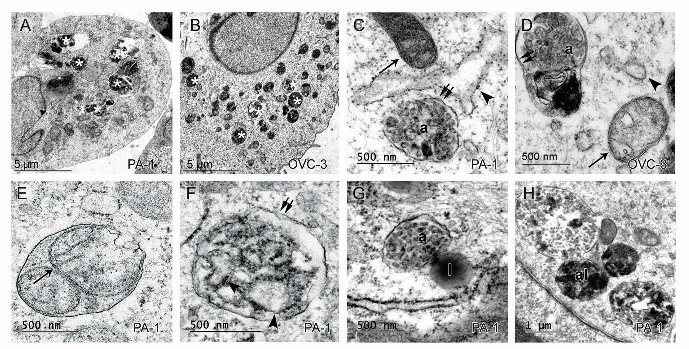
Fig Monitoring autophagy by TEM
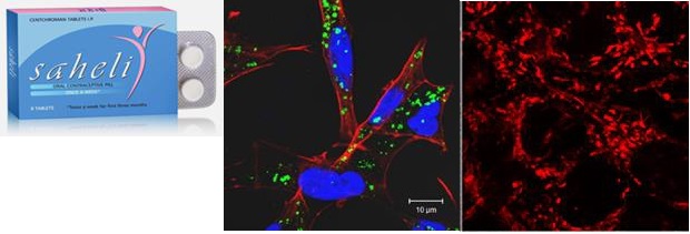
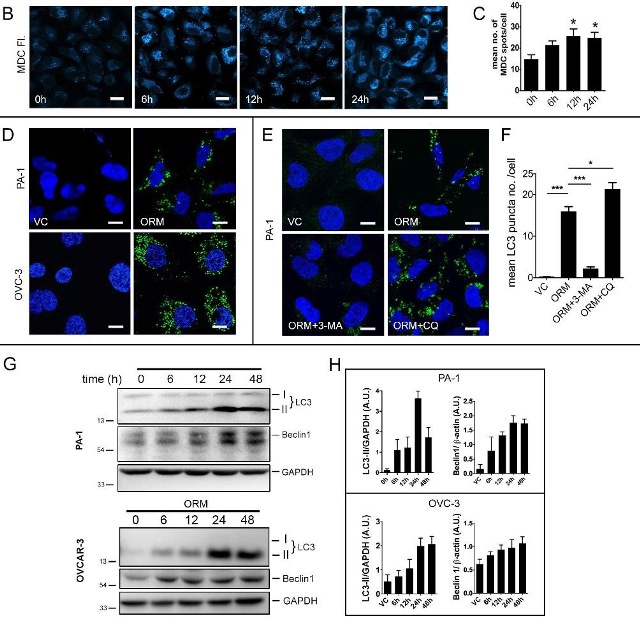
Sci Rep. 2018 Feb 2;8(1):2303.
Anti-proliferative activity of natural compounds and their modes of action are also being investigated.
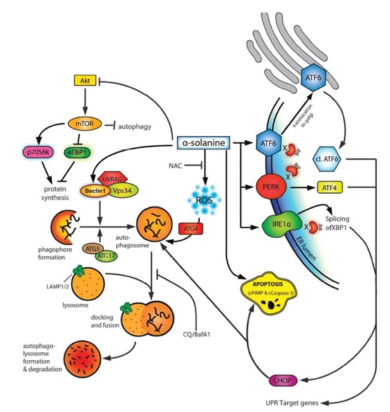
Fig. Proposed mechanism of α-solanine-induced cell death. Cell Death and Disease (2015)
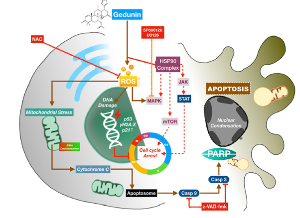
Fig. Schematic representation of proposed mechanism of action of Gedunin in ovarian cancer cells (Apoptosis. 2020)
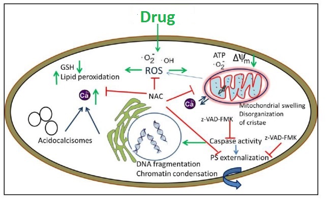
2. Evaluation of potential anti-leishmanial agents and investigating their mechanism of action
In our ongoing efforts to identify and understand the mode of action of new and effective leishmanicidal agents, several synthetic and natural compounds
are currently being evaluated in our laboratory. Molecular machinery of cell death in Leishmania differs considerably with metazoan apoptosis and is not
well understood. Studies addressing these aspects may aid to not only determine the significance of these molecules in the life cycle of these organisms
but also to provide tools to design drugs against Leishmaniasis. Our strategy is to target unique organelles/essential metabolic pathways with steps
differing from those found in mammalian hosts – mitochondrion (Antimicrobial Agents & Chemotherapy 2014 Oct;58(10):5916-28); Ergosterol synthesis pathway
(Apoptosis 2018 Aug;23(7-8):420-435); Trypanothione pathway (Apoptosis 2016 Aug;21(8):941-53).
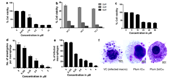
Fig. Evaluation of anti-proliferative activity demonstrating dose and time dependent effects of the compound in both forms of the parasite.
Fig.
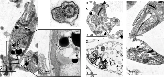
Fig. TEM analysis revealing fine structure alterations in Leishmania promastigotes providing valuable insights into the mechanism of action of
anti-leishmanial agents.
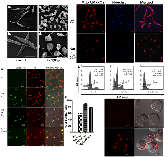
Fig. SEM micrographs demonstrating morphological effects of the compound; Visualizing mitochondrial depolarisation using confocal microscopy; determination
of apoptotic cell death by TUNEL assay; Cell cycle analysis by Flow cytometry; mitochondrial superoxide formation in intracellular amastigotes
3.Development of novel fluorescent probes for cell imaging applications:
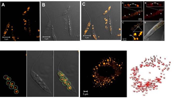
Fluorescent Probe for Selective Staining and Quantification of Intracellular Lipid Droplets.
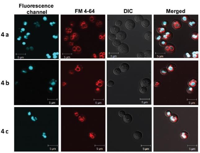
Fluorescent probes for selective staining of vacuoles
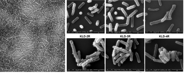
4. Other collaborative projects include EM characterization of drug delivery systems and biological nanostructures and macromolecular complexes.
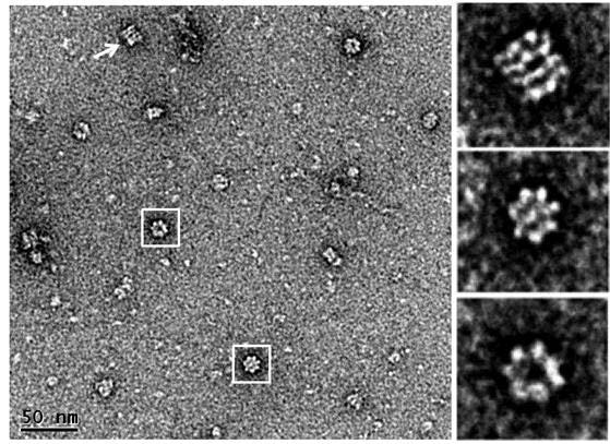
Fig. TEM micrograph revealing structural organization of the homo-oligomeric complex of LdTCP1c, revealing sevenfold symmetry (boxed). A side view of the
complex (arrow) reveals the double ring structure. The images/1614_kalyan on the right show different views of the complex. FEBS J. 2015 Dec;282(23):4607-19
Drug Delivery systems/Formulations:
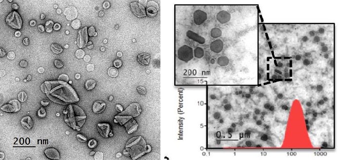
Rutin phospholipid complexes Nano Liquid Crystalline Particles
RSC Adv., 2016. ACS Appl. Mater. Interfaces, 2018
Assessing Self-sssembling properties of peptides and their anti-microbial effects by EM Biomaterials, 2015