Lymphatic filariasis - An introduction
Lymphatic filariasis (LF) is an incapacitating and disfiguring neglected tropical disease (NTD) caused by Wuchereria bancrofti, Brugia malayi and B. timori. Presently 886 million people in 52 countries are threatened by LF and approximately 65% of these live in the south-east Asia region including India, where people in 18 states and 6 union territories are at risk of infection. Elephantiasis, Lymphoedema and Hydrocele are the major pathological outcomes of LF (Fig 1).
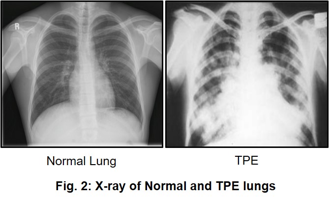
Besides this, Tropical Pulmonary Eosinophiila (TPE) is a rare and serious inflammatory disorder of the lungs that is seen in a small minority of patients infected with filarial parasites. Untreated TPE can lead to lung fibrosis and even death of patients, making TPE the only filarial complication that results in mortality (Fig 2).
LF causes untold pain, social and mental stigma, and impairment of the body’s ability to fight infection leading to significant economic loss, reduced productivity and psychosocial effects on the infected individual and the community. Depending on the geographical region, several species of mosquitoes (Aedes, Anopheles, Culex etc.) serve as vectors of LF. Currently available mainstay drugs viz. Diethylcarbamazine (DEC), Ivermectin (IVM) and Albendazole (ALB) are mainly microfilaricidal in nature and exhibit only modest adulticidal activity which means that they are effective in stopping transmission of the disease, but do not kill the adult worm. Though newer strategies based on targeting the Wolbachia endosymbiont which is essential for worm fertility and survival are being studied, vaccine for any filarial parasite is not yet a reality.
Lab research
The current focus of the lab is cutting edge research in understanding the immunobiology of Lymphatic Filariasis as well as Drug target and drug discovery of new anti-filarial compounds. Basic research in the lab revolves around three main areas of interest which are briefly discussed below.
1. Role of host immune cells during Lymphatic filariasis:
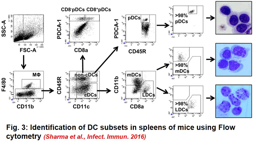
Parasites have evolved many diverse and novel strategies to evade the host immune response which includes dampening of the host’s pro-inflammatory cytokine response, attenuating the functions of innate immune cells, generating regulatory T cells and impairing the activation, maturation and functions of host macrophages and dendritic cells (DCs). Though few reports have looked into the role of primary host DCs during the very early stages of filarial infection, DC subset specific studies are not only lacking but it is also not clear as to what extent these subsets are affected during the early stages of filarial infection. One of the main focus in the lab is therefore to identify DC subsets in the host lymphoid organs post LF infection, and study their functional impairment. We have successfully identified different DC subsets (Myeloid, Lymphoid and plasmacytoid DCs) in spleens and mLNs of infected mice (Fig. 3) and showed that these subsets have impaired antigen uptake and presentation capacity that leads to selective attenuation of their T cell proliferation capacity post infection with infective larvae of the parasite.
In addition to this, finding out the reasons behind alternative activation of host macrophages and subsequent impaired priming of the host adaptive immune response is an important aspect of our research work.
2. Role of eosinophils during Tropical Pulmonary Eosinophilia (TPE):
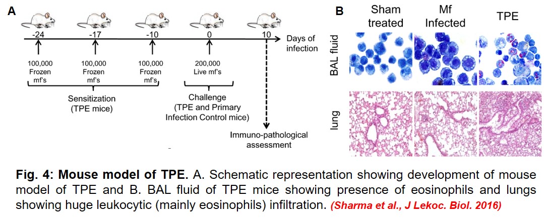
Eosinophils are centrally involved in the pathology of TPE. Lung lavage and lung biopsies of TPE patients show dominance of eosinophils whose numbers are correlated with severity of pathological manifestations of TPE. However, eosinophils are very fragile cells, their low numbers and dual nature (protective and pathological) makes their isolation extremely difficult. Interestingly, it is this behavior of eosinophils that has made the immunological etiology of TPE very confusing and complicated. Our lab has recently developed a mouse model of TPE that exhibits cardinal features of human TPE pathogenesis (Fig. 4).
Using Flow cytometry, we have successfully identified and sorted eosinophils to high purity, and have identified some key genes and proteins that are differentially expressed during TPE. Biochemical and immunological characterization of these genes and proteins will help us to delineate their role during TPE and also ascertain whether targeting them will mitigate TPE associated symptoms in the lungs.
3. Immunogenicity and protective efficacy of Filarial/Wolbachial antigens:
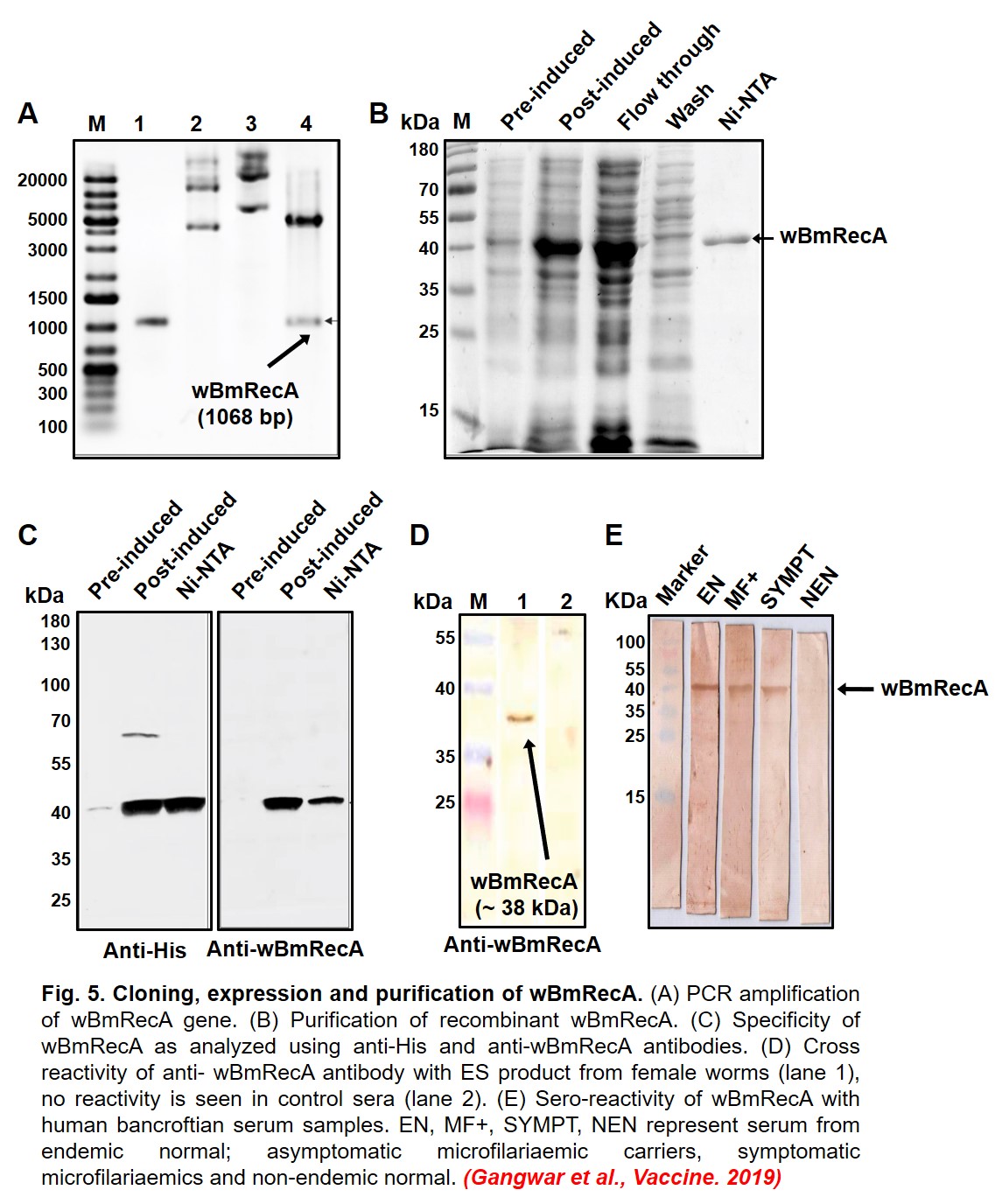
Wolbachia is an endosymbiotic intracellular bacterium of the filarial nematode that helps in their growth and development, regulates fecundity in female worms and contributes to the immunopathogenesis of the disease. However, genes and proteins of Wolbachia that may act as putative vaccine candidates are not known. Our research group has identified couple of interesting candidates among them Recombinase A from Wolbachia (wBmRecA) that are highly immunogenic and thus are promising vaccine candidates against filariasis (Fig 5). The lab is also interested in investigating the immunogenicity of various Brugia malayi antigens that modulate the protective host immune response during LF infection.
Drug target and drug discovery: The lab has an associated insectarium where Aedes aegypti are reared and bred and used for cyclic maintenance of infection in small rodents viz. Mastomys coucha and Meriones unguiculatus that are permissive to filarial infection (Fig. 6). The lab routinely carries out screening of synthetic and natural products for evaluation of their anti-filarial activity using both in vitro and in vivo approaches.